Skull base & pituitary surgery — MCW / Froedtert

Nathan T.
Zwagerman, MD, FAANS, FACS

Neurosurgeon specializing in the cranial base — endoscopic endonasal approaches to the pituitary gland, transorbital and mini-orbitozygomatic corridors, and lateral skull base surgery for acoustic neuroma and semicircular canal dehiscence. Territory where anatomy leaves the smallest margin for error, and where a coordinated team matters as much as the surgeon.
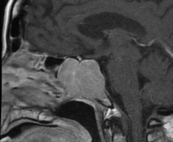
entry sella turcica
APPROACHtransnasal · transsphenoidal · endoscopic
TARGETsella turcica — pituitary gland
DISCIPLINEneurosurgery + otolaryngology, combined
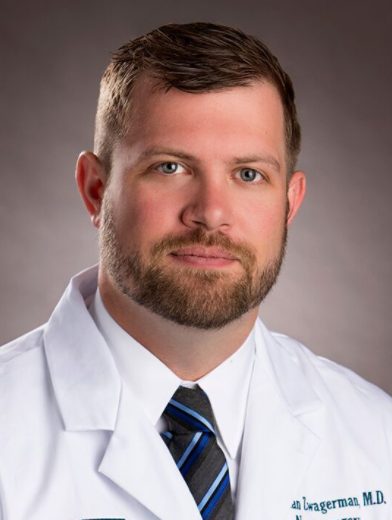
Portrait of Dr. Nathan T. Zwagerman
Background

Academic neurosurgeon, high-volume operator.

Dr. Zwagerman is an Associate Professor of Neurosurgery, Otolaryngology, and Surgery at the Medical College of Wisconsin and Froedtert Hospital. His practice centers on pituitary and skull base pathology, with an emphasis on minimally invasive endoscopic endonasal technique performed in close partnership with rhinology colleagues.

  • ClinicalDirector of Pituitary and Skull Base Surgery, Medical College of Wisconsin / Froedtert Hospital.
  • EducationProgram Director, Neurosurgery Residency and Skull Base Fellowship.
  • AdministrationVice Chair of Administration, Department of Neurosurgery.
  • SocietyBoard leadership, North American Skull Base Society (NASBS).
Clinical focus

Precision where the brain meets the face.

01

Pituitary adenomas

Functioning and nonfunctioning tumors of the pituitary gland, evaluated jointly with endocrinology.

02

Skull base tumors

Craniopharyngioma, meningioma, and other lesions of the anterior and central cranial base.

03

Endoscopic endonasal surgery

Transnasal, transsphenoidal approaches performed without external incisions where anatomy allows.

04

Cranial base reconstruction

Multilayer closure technique aimed at minimizing CSF leak and reoperation.

05

Transorbital & mini-orbitozygomatic approaches

Minimally invasive eyelid or brow corridors to the orbital apex, anterior, and middle fossa — an alternative to larger craniotomies for select lesions.

06

Lateral skull base

Acoustic neuroma (vestibular schwannoma) microsurgery and semicircular canal dehiscence repair, performed jointly with neurotology.

Imaging & the operating room

Selected case imaging.

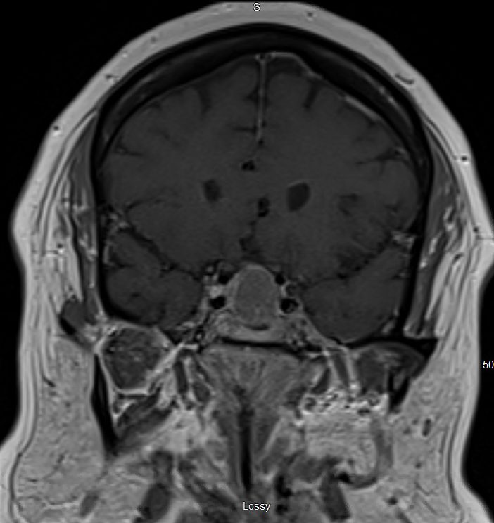
De-identified case examples, shared with consent. Labels below are a starting description — swap in exact diagnoses and case details as you see fit.

Sagittal MRI of a sellar region mass
Sagittal MRI

Pituitary / sellar region mass

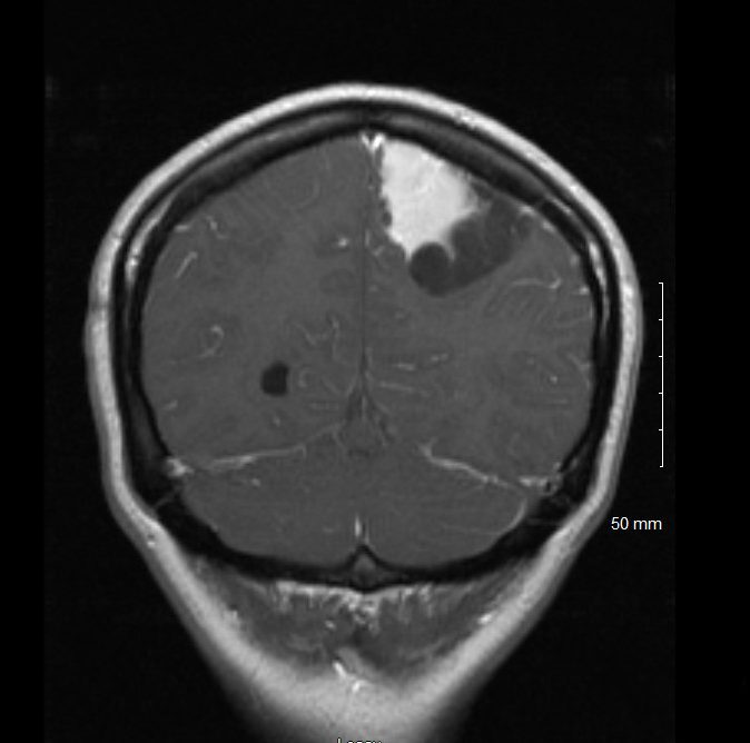
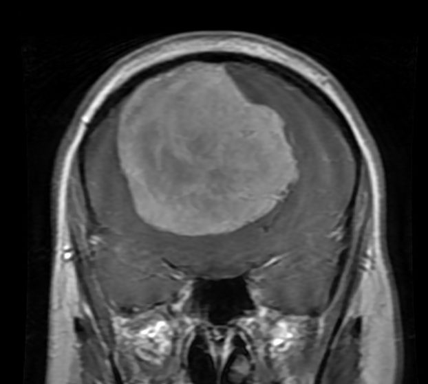
Coronal MRI of a large convexity mass
Coronal MRI

Convexity meningioma

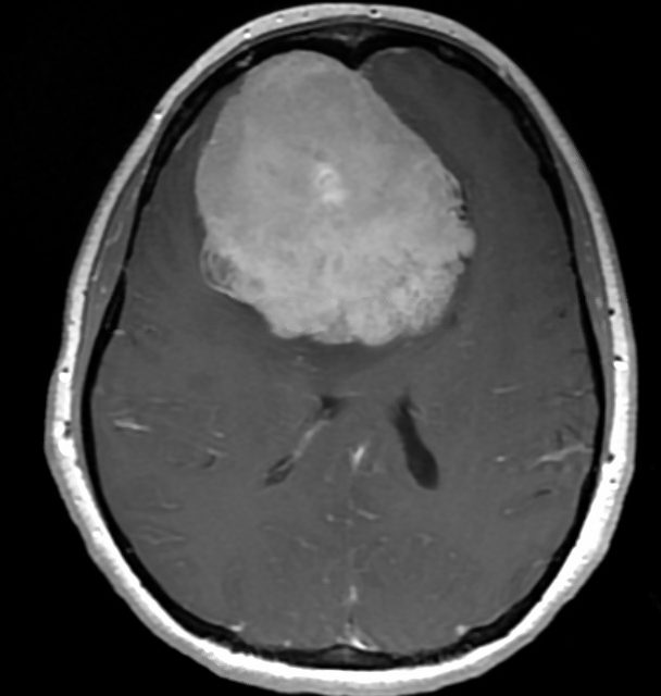
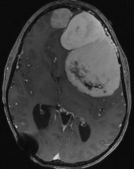
Axial MRI of a large convexity mass
Axial MRI

Convexity meningioma

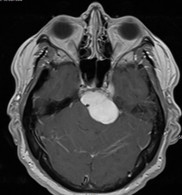
Axial MRI of a petroclival region lesion
Axial MRI

Petroclival lesion

Axial MRI of a lateral skull base mass
Axial MRI

Lateral skull base tumor

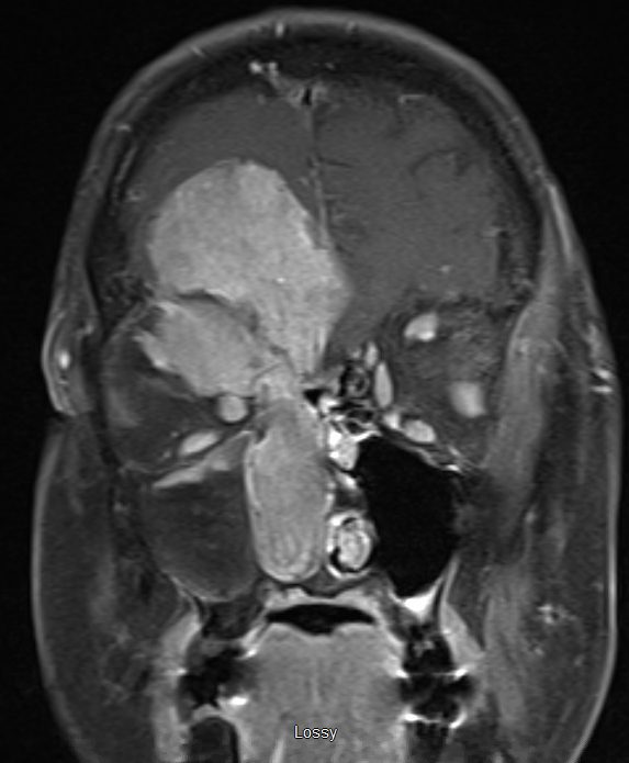
Coronal MRI of a skull base mass
Coronal MRI

Skull base tumor

Axial MRI of a tumor with hemorrhage
Axial MRI

Tumor with hemorrhage

Coronal MRI of a tumor with hemorrhage
Coronal MRI

Tumor with hemorrhage

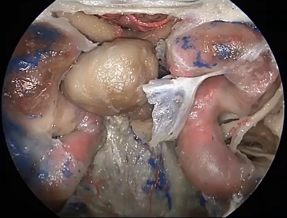
Intraoperative endoscopic view during skull base tumor surgery
Intraoperative

Endoscopic operative view

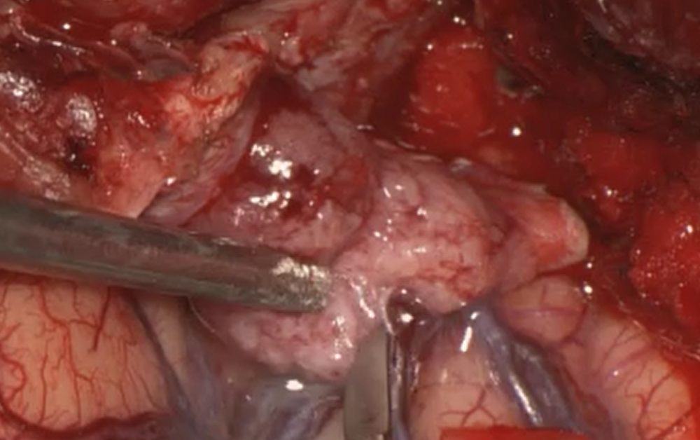
Intraoperative microscopic view during skull base tumor dissection
Intraoperative

Microscopic tumor dissection

For patients

Understanding a skull base diagnosis.

A skull base or pituitary diagnosis is unfamiliar territory for almost every patient who receives one. These notes are meant as a starting point for the conversation, not a substitute for it — every plan is built around your imaging, your symptoms, and your goals.

Pituitary

What the pituitary gland does

The pituitary sits at the base of the brain and regulates hormones that affect growth, metabolism, stress response, and reproduction. Tumors here can be "functioning" (producing excess hormone) or "nonfunctioning" (found because of size or incidental imaging), and evaluation is done jointly with endocrinology.

Symptoms

What often prompts a workup

Headaches, vision changes, hormone irregularities, or a mass found incidentally on imaging obtained for another reason are the most common paths to a skull base referral. Not every finding needs surgery — some are monitored with serial imaging.

Hearing & balance

Acoustic neuroma & dehiscence

Gradual hearing loss, tinnitus, or unsteadiness can point to a lateral skull base process such as acoustic neuroma or semicircular canal dehiscence. These are evaluated with dedicated imaging and audiometric/vestibular testing before any decision about surgery.

What to expect

Evaluation & second opinions

A first visit typically reviews prior imaging, orders any additional studies needed, and lays out the realistic range of options — observation, medical management, or surgery — before any decision is made. Second opinions on existing imaging are welcome.

Research & academic leadership

Translational work grounded in the operating room.

Biomarkers

O-GlcNAcylation in pituitary tumors

Long-standing translational collaboration with Dr. Stephanie Olivier-Van Stichelen (MCW Biochemistry) studying O-GlcNAcylation in pituitary adenomas and corticotroph tumors, supported by an NASBS research grant.

Grant-funded

Risk stratification for nonfunctioning adenomas

AHW Endowment-funded project developing biomarker-based risk stratification for nonfunctioning pituitary adenomas, drawing on the MCW tumor biobank.

Multi-site

Trial leadership & consortia

Site Principal Investigator for the BOOST TBI Trial and TRACK-NET, and a participating site in the RAPID craniopharyngioma consortium.

73+
Peer-reviewed publications
3
Active multi-site trials / consortia
NASBS
Board-level leadership
MCW
Residency & fellowship direction
Publications

73+ peer-reviewed papers across skull base practice.

A full, current bibliography is maintained externally rather than duplicated here.

Cluster

Pituitary biomarkers & O-GlcNAcylation

Work on O-GlcNAcylation signaling in pituitary adenomas and corticotroph tumors, and biomarker-based risk stratification for nonfunctioning adenomas.

Cluster

Skull base surgical outcomes

Outcomes and technique papers spanning endoscopic endonasal, transorbital, and lateral skull base approaches.

Cluster

Trauma & craniopharyngioma

Contributions tied to the BOOST TBI Trial, TRACK-NET, and the RAPID craniopharyngioma consortium.

Speaking, media & video

Selected engagements and recordings.

NASBS

Petroclival lesions — session faculty

North American Skull Base Society annual meeting.

MCW

Neurosurgery grand rounds

Regular participation in departmental grand rounds programming at the Medical College of Wisconsin.

[add additional talks, podcasts, or press]

Placeholder row — extend this list as engagements are booked or published.

Global health

Building surgical capacity beyond Milwaukee.

Dhulikhel Hospital, Nepal

An ongoing partnership, established in 2023, focused on building sustainable neurosurgical and skull base capacity through training and shared casework.

WINS Program

A cross-departmental collaboration between Neurosurgery and Medicine that has drawn national recognition for its model of coordinated, team-based care.

Contact

For referring physicians and patients.

Referring physicians

For expedited review of pituitary and skull base cases, reach the office directly or route imaging through the MCW referral line.

InstitutionMedical College of Wisconsin / Froedtert Hospital
Referral line414.805.5400
Secure imaging transfer[add link]
Froedtert profilefroedtert.com ↗
MCW profilemcw.edu ↗

Patients

New patient consultations for pituitary and skull base conditions are coordinated through the Froedtert & MCW neurosurgery clinic.

ClinicFroedtert & MCW Neurosurgery
Scheduling414.805.5400
LocationMilwaukee, WI
Froedtert profilefroedtert.com ↗
MCW profilemcw.edu ↗